PRJ002AiptasiaMM Knockdown Sampling Protocol
Objective
PRJ002 aims to reduce the microbiome of Exaiptasia pallida. We tested and compared 2 antibiotic solutions plus a control population. This post serves to establish the sampling protocol used across all our sampling timepoints following the initial Start point. See T0 Notes
Sampling Time Points
Anemones were primed in FSW for 1 month at which point 5 experimental (combined ABS1 and ABS2) and 5 control anemones were sampled (T1). Anemones were then treated for 3 weeks with their respective ABS solutions. Samples were taken weekly ( 5/ condition) during this time period (T2-4) Finally Samples will be taken 1 and 2 weeks post treatment with ABS treated individuals being kept in Filtered Autoclaved Artificial Seawater(FAASW).
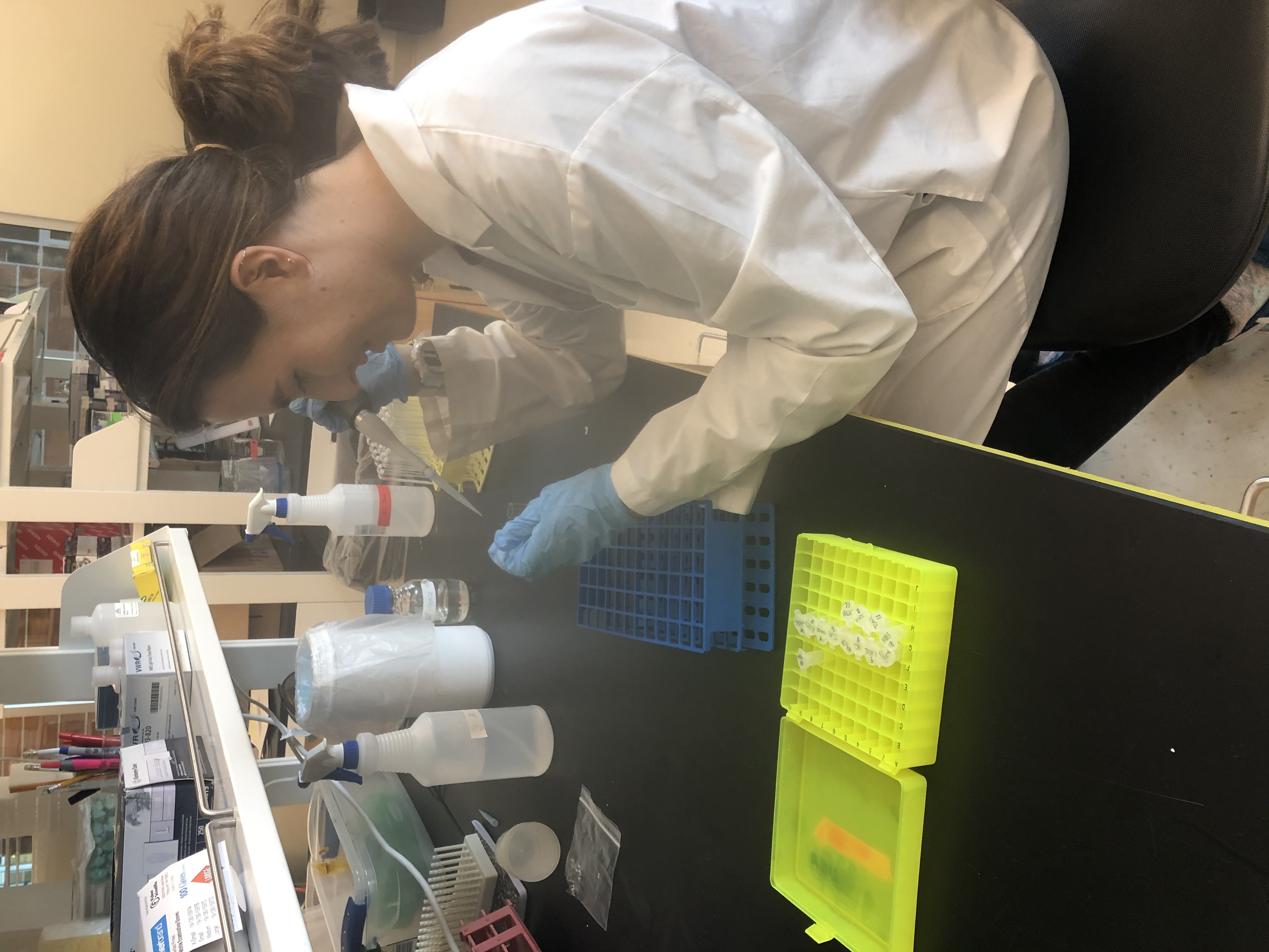
Sampling protocol.
5 anemones/ condition were moved to new plates and allowed 24 hours of recovery outside of ABS solution prior to sampling. Anemones were picked into 1.5ml tubes containing 250ul of FAASW and homogenized using a pestle motor until no identifiable anemone remained. 300ul of FAASW added to the tube and samples were vortexed for 5s to mix. Samples were then aliquoted off for 3 different sub samples.
DNA Sampling:
250 ul of sample was added to an beadbeating tube with Lysing matrix B and D and 500ul of DNA/RNA Shield. samples were bead beat at 8 m/s for two cycles of 60 seconds with a five minute pause between cycles, then frozen at -80C until Extraction.
CFU counts
70ul of homogenate was aliquoted for serial dilutions. Homogenate was diluted 10, 100 and 1000 fold. 50 ul of homogenate/dilution were plated on Marine Agar plates and spread using plating beads. plates were allowed to incubate for 24 hours at 28C at which point CFU counts were obtained.
Flow Cytometry and Protein content
20ul of 0.1% SDS solution were added to the remaining aprox. 180ul of sample homogenate ( final conc. 0.01%SDS) and frozen for downstream processing
Physiology plates
6 samples / condition were set aside for physiology metrics to be taken at each time point throughout the experiment.
to monitor morphological changes
anemones were transferred to a sterile petri dish to measure wet weight
a Keyence BZX710 inverted microscope was used to image individual at each time point and photos of whole plates were also taken with on an iphone
Finally Pedal lacerates were counted weekly.. samples were moved accross plates once per week during the priming phase and pedal lacerates were discarded, but pedal lacerates from the knockdown phase were kept for the duration of the experiment.